
Spermerz Logo
Back to Programs
Clinical Research & Insights
Uterine Fibroids Care & Clinical Support
Medically Reviewed by Tabib Dr. Sajid Nawaz (NCH-78420-PK)
Clinical Verification: August 2026

Uterine Fibroids (Leiomyomas) & Abnormal Bleeding: FIGO Classification, MRI Reports, and Management

Comprehensive guide to uterine fibroids (leiomyomas) and abnormal uterine bleeding: FIGO staging, reading MRI reports, red degeneration, and myomectomy options.

Uterine Fibroids (Leiomyomas) & Abnormal Bleeding: FIGO Classification, MRI Reports, and Management

1. Definition and Cellular Pathogenesis

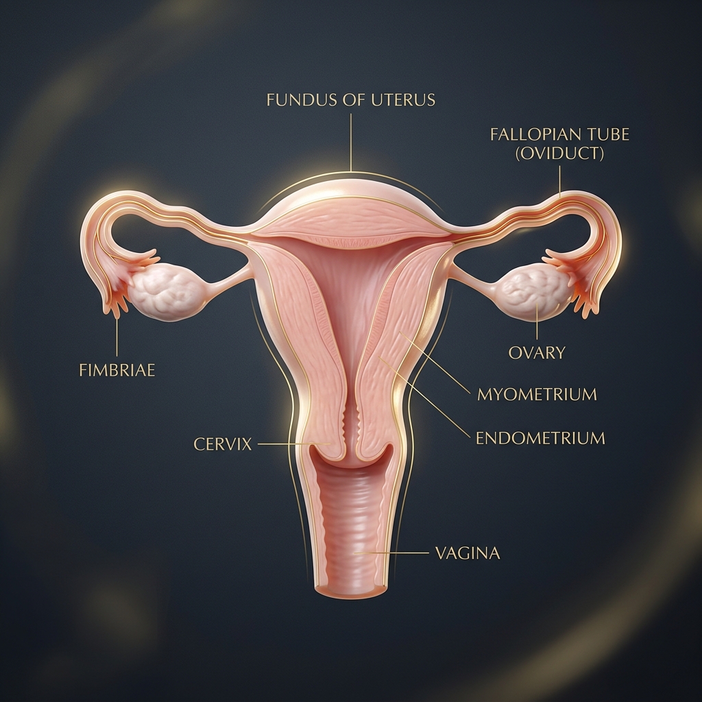
Uterine Leiomyomas (Fibroids) are benign monoclonal smooth muscle neoplasms arising from the myometrium of the uterus. They are the most common pelvic tumor in women of reproductive age, present in up to 70–80% of women by age 50. Growth is highly steroid-dependent, driven by estrogen and progesterone, overexpressing both estrogen receptors (ER-α) and progesterone receptors (PR-A, PR-B). They are characterized by excessive deposition of disordered extracellular matrix (ECM) rich in collagen, fibronectin, and proteoglycans.

2. The FIGO PALM-COEIN & Fibroid Classification System

The International Federation of Gynecology and Obstetrics (FIGO) categorizes the causes of Abnormal Uterine Bleeding (AUB) and classifies fibroids based on their anatomical depth within the uterine wall:

                        FIGO FIBROID SUBTYPES (0 to 8)
                                      β”‚
     β”Œβ”€β”€β”€β”€β”€β”€β”€β”€β”€β”€β”€β”€β”€β”€β”€β”€β”€β”€β”€β”€β”€β”€β”€β”€β”€β”€β”€β”€β”€β”€β”€β”€β”Όβ”€β”€β”€β”€β”€β”€β”€β”€β”€β”€β”€β”€β”€β”€β”€β”€β”€β”€β”€β”€β”€β”€β”€β”€β”€β”€β”€β”€β”€β”€β”€β”€β”
     β–Ό                                β–Ό                                β–Ό
SUBMUCOSAL (Types 0-2)          INTRAMURAL (Types 3-5)           SUBSEROSAL (Types 6-7)
β”œβ”€β”€ 0: Pedunculated intracavitaryβ”œβ”€β”€ 3: 100% myometrial, contacts β”œβ”€β”€ 6: Subserosal ≥ 50%
β”œβ”€β”€ 1: < 50% intramural              endometrium                 β”œβ”€β”€ 7: Pedunculated subserosal
└── 2: ≥ 50% intramural         β”œβ”€β”€ 4: 100% intramural           └── 8: Other (Cervical / Parasitic)
                                └── 5: Subserosal < 50%
Type Classification Anatomical Relationship Clinical Impact
Type 0 Submucosal Pedunculated within the endometrial cavity Severe menorrhagia, prevents embryo implantation
Type 1 Submucosal Intramural component < 50% Profuse bleeding, recurrent miscarriage
Type 2 Submucosal Intramural component ≥ 50% Heavy bleeding, requires combined surgical approach
Type 3 Intramural 100% intramural; touches the endometrium Heavy flow, dysmenorrhea
Type 4 Intramural Fully embedded within myometrium Dysmenorrhea, bulk-related pelvic pain

3. Clinical Symptoms & Red Degeneration

  • Abnormal Uterine Bleeding (AUB / Menorrhagia): Prolonged menses (> 8 days), passing large blood clots, flooding through sanitary products, resulting in secondary severe iron deficiency anemia.
  • Pelvic Pressure & Bulk Symptoms: Sensation of lower pelvic heaviness, increased abdominal girth, bladder tenesmus, and frequent nighttime urination.
  • Acute Red (Carneous) Degeneration: Occurs primarily during pregnancy or rapid hormonal stimulation when the fibroid outgrows its blood supply, causing acute ischemic infarction, severe localized pain, localized peritoneal signs, and low-grade fever.

4. How to Read Your Pelvic Ultrasound & MRI Reports

========================================================================================
PELVIC ULTRASOUND & MRI SCAN INTERPRETATION FOR UTERINE FIBROIDS

1. ULTRASOUND CHARACTERISTICS
β”œβ”€ Classic Benign Fibroid: Well-circumscribed, hypoechoic, solid mass with edge shadowing
β”‚  and posterior acoustic attenuation ("venetian blind" shadowing).
β”œβ”€ Degenerating Fibroid: Anechoic/cystic internal spaces, fluid levels, calcific rims.
└─ Endometrial Line Distortion: Submucosal displacement into cavity directly visualized.
2. MAGNETIC RESONANCE IMAGING (MRI) INTERPRETATION (Gold Standard for Surgical Mapping)
β”œβ”€ T1-Weighted Imaging: Benign leiomyomas appear isointense to normal myometrium.
β”‚  * Red Degeneration: Displays a characteristic hyperintense peripheral rim on T1.
β”œβ”€ T2-Weighted Imaging: Non-degenerating fibroids appear sharply demarcated and HYPOINTENSE
β”‚  (very dark), matching fibrous smooth muscle bundles.
β”‚  * High T2 Signal Intensity (Bright): Indicates cellular leiomyoma or myxoid degeneration.
β”œβ”€ Post-Contrast T1 Gadolinium Enhancement:
β”‚  * Uniform Enhancement: Viable, highly vascularized active fibroid tissue.
β”‚  * Non-Enhancing Regions: Avascular necrotic tissue (infarcted/hyalinized).
└─ Leiomyosarcoma Suspicion: Ill-defined borders, rapid post-menopausal growth, and restricted diffusion on DWI.
========================================================================================

5. Medical, Interventional, and Surgical Management

                       TREATMENT OPTIONS FOR FIBROIDS
                                     β”‚
 β”Œβ”€β”€β”€β”€β”€β”€β”€β”€β”€β”€β”€β”€β”€β”€β”€β”€β”€β”€β”€β”€β”€β”€β”€β”€β”€β”€β”€β”€β”€β”€β”€β”€β”€β”€β”€β”Όβ”€β”€β”€β”€β”€β”€β”€β”€β”€β”€β”€β”€β”€β”€β”€β”€β”€β”€β”€β”€β”€β”€β”€β”€β”€β”€β”€β”€β”€β”€β”€β”€β”€β”€β”€β”
 β–Ό                                   β–Ό                                   β–Ό
PHARMACOLOGICAL                     INTERVENTIONAL                      SURGICAL
β”œβ”€β”€ GnRH Antagonists                β”œβ”€β”€ Uterine Fibroid Embolization   β”œβ”€β”€ Hysteroscopic Myomectomy
β”‚   (Relugolix, Lupron)                 (UFE / UAE)                    β”œβ”€β”€ Laparoscopic Myomectomy
β”œβ”€β”€ Tranexamic Acid (Bleeding)      └── Focused Ultrasound Surgery     β”œβ”€β”€ Abdominal Open Myomectomy
└── Progestin IUD (Mirena)              (HIFU)                         └── Total / Subtotal Hysterectomy
  1. Medical Therapies: Oral GnRH Antagonists (Relugolix / Elagolix combined with add-back therapy) to control bleeding and shrink fibroids; Tranexamic Acid to reduce heavy blood loss during menses.
  2. Uterine Fibroid Embolization (UFE/UAE): Interventional radiologist injects microsphere particles into both uterine arteries, cutting off blood supply to all fibroids.
  3. Surgical Interventions: Hysteroscopic Myomectomy (transcervical resectoscopic loop excision for submucosal types); Laparoscopic / Robotic Myomectomy (for subserosal and intramural fibroids while preserving fertility); Hysterectomy (definitive cure).

🚨 Emergency Red Flags β€” Seek Immediate Medical Evaluation If:

  • Bleeding heavily enough to soak through two or more maxi pads per hour for two consecutive hours.
  • Sudden onset of severe, unmanageable pelvic pain accompanied by nausea and fever (pedunculated torsion or red degeneration).
  • Severe dizziness, shortness of breath, or pale skin (acute severe anemia).
Discuss this research privately with our men's health specialist.
Discuss this article on WhatsApp
100% Private & Confidential

Ready to solve this concern permanently?

General information is helpful, but every body and history is unique. Get custom herbal support and a tailored plan today.

Topics & Tags

#uterine-fibroids #leiomyoma #figo-classification #myomectomy